How Minimally Invasive Surgery Got Melanie Back on Her Feet
Melanie Goshgarian, 48, lives in the seacoast town of Scituate, Massachusetts. After moving there six years ago, she enjoyed walking along the nearby beach each morning. Eventually, though, that became impossible due to extreme pain in her right foot.
By the time she saw Mass General Brigham Orthopedics and Sports Medicine foot and ankle surgeon Christopher Miller, MD, her foot was in bad shape. Because of a complex immunological condition that she has, finding a safe and effective treatment option would prove to be complicated.
According to Dr. Miller, Melanie had likely had an underlying foot deformity for many years. Then she developed a bunion, a painful bump that forms when the first metatarsal bone (at the base of the big toe) shifts out of place.
As it got worse, the bunion pushed Melanie’s big toe toward her second toe. Over time, that crowding put extra stress on the second toe, forcing it to buckle upward at the joint. The result was an aching hammertoe.
Her foot troubles came to a head when Melanie stubbed her right second toe on the foot of a bench. She figured it was just a sprain. But the impact had weakened the ligaments supporting the toe, making an already unstable foot even more painful.
Over the next six months, Melanie dealt with bruising and worsening pain. She toughed it out as best she could. Then came a big snowstorm.
“While lifting trash bins over a snowbank, I felt something shift in my foot,” she recalled. “The pain was bad enough that I had to get a ride to Mass General Brigham Express Care because I couldn’t even press on the gas pedal or brakes in the car.”
It was the first step in a care journey that led to successful surgery to repair her foot.
Woman smiling while cuddling a dog on a couch.
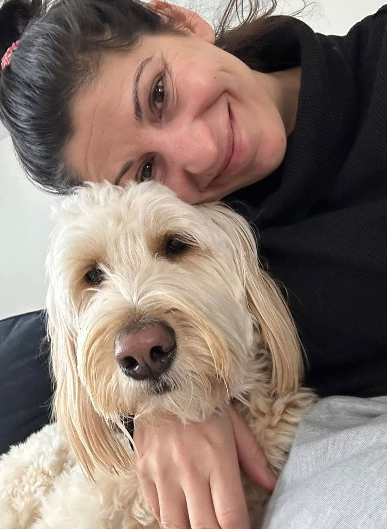
Melanie and her dog
A challenging case
After evaluating her foot, a nurse practitioner at Express Care referred Melanie to Dr. Miller’s office. At her first visit a few days later, Melanie had an MRI and saw physician assistant Brianna Whitehouse, PA-C. Brianna suspected surgery would be needed to repair the foot.
A week later, Melanie saw Dr. Miller of the Foot and Ankle Care program for the first time. He recognized right away that this would be a challenging case.
A surgeon would typically address an array of foot problems like Melanie’s with metal screw fixation. This technique involves using stainless steel or titanium screws to stabilize bones and keep them in the correct position. Due to a significant metal allergy and history of severe allergic reactions, however, metal hardware would not be an option for Melanie.
“At that point, we were trying to figure out how we could correct her deformity and treat her pain, all while dealing with this real limitation,” Dr. Miller said.
Dr. Miller’s solution was a minimally invasive surgical technique that is common in France and Spain but very rare in the United States. He traveled to Paris almost a decade ago to learn about it from a colleague.
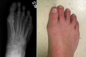
A photo and X-ray show Melanie’s bunion, where the big toe had shifted toward the smaller toes and created a painful bump on the inside of her foot.
Minimally invasive scarf osteotomy
In considering Melanie’s case, Dr. Miller thought that scarf osteotomy would be a good fit. This procedure involves cutting a long, Z-shaped pattern to divide the first metatarsal bone into two pieces. The pieces are then shifted to correct the toe’s alignment and secured in place like a jigsaw puzzle. Lastly, the bony bump is shaved to achieve a smoother profile on the side of the foot.
Surgeons usually perform a scarf osteotomy through a larger incision (open surgery) and use metal screws to hold the bones in place while they heal. Dr. Miller—an expert in minimally invasive surgery for the foot and ankle—had a different approach in mind.
He proposed doing a minimally invasive scarf osteotomy. This would require making only a 5 millimeter cut, through which he would insert tiny, specialized tools to cut the bone.
Using a minimally invasive technique would:
- Minimize disruption to surrounding tissue.
- Reduce the risk of wound complications.
- Minimize scarring and postoperative pain.
- Speed a return to walking and overall recovery.
“By positioning each piece of the metatarsal bone next to the other, I could maximize the amount of contact so that the bone could heal on its own. That would make for an intrinsically stable solution,” Dr. Miller said. “Plus, I wouldn’t have to use screws—a necessity given Melanie’s metal allergy.”
After talking through the benefits and risks with Dr. Miller, Melanie felt comfortable moving forward with minimally invasive scarf osteotomy.
“Dr. Miller was very easy to talk to and didn’t try to push anything on me, which I’ve felt with other surgeons,” she said. “He and everyone on his team, from Brianna to the front desk staff, were compassionate and understanding.”
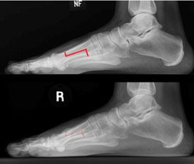
X-rays show where Dr. Miller planned to cut and realign the bone, and how the bone healed afterward while staying in the correct position.
Enjoying a rapid recovery
The one-hour operation took place in the morning. Melanie woke up afterwards to a nice surprise: Since the surgery had gone so smoothly, she would be able to walk as soon as she regained feeling in her right foot—much sooner than Dr. Miller had anticipated.
Melanie returned home early the same afternoon wearing an air cast boot on the foot. Since no screws were used, she had been fitted with special dressings to maintain the bony alignment.
“We wet the dressings and then fold them around the toe,” Dr. Miller said. “As they dry, they almost turn into little casts to hold the bones in place.”

X-rays taken during surgery show how Dr. Miller used a small surgical tool to cut and realign the bone, helping correct the shape of Melanie’s foot.
Two weeks later, Melanie had a follow-up visit with Dr. Miller. As he rebandaged her toe, she glanced at her foot. “I was like, ‘Where’s the scar?’ There was nothing but a tiny little ‘X,’ which has since faded away,” she said.
By her four-week visit with Dr. Miller, Melanie was getting around pretty well. She didn’t need pain medication anymore—just ice and rest. About 10 days after that, she was able to drive to Dr. Miller’s office for an appointment. X-rays at each visit confirmed her foot was healing properly.
In between physical therapy sessions with a local provider, Melanie has gradually gotten back to normal activities like walking, yoga, Pilates, kayaking, and paddleboarding. With the surgery six months behind her, she couldn’t be happier with the results.
“Everything in my foot is definitely in the right place,” she said. “I get some swelling and pain if I overdo it, but otherwise those aren’t an issue. I’d been really limited, and I’m able to do more now than I was before.”
As an access coordinator at Brigham and Women’s Hospital, Melanie regularly interacts with patients who worry that their health problems are too complex to be treated.
“I used to feel that way, too,” she said. “But this was well-coordinated, timely care, with a very successful outcome. I hope my story gives other patients in my shoes some reassurance.”

At Melanie’s two-week follow-up visit, her foot was healing well and her big toe was back in better alignment.
Learn about foot and ankle care
Learn more about foot and ankle care