AI Advancements Make the Leap into 3D Pathology Possible
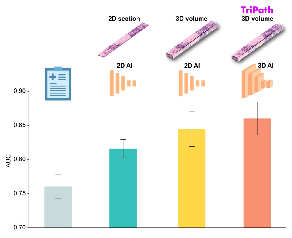
Researchers developed Tripath to bridge computational gaps to process 3D tissue and predict outcomes based on 3D morphological features.
Cancer recurrence models trained on 3D tissue volumes outperformed models trained on 2D tissue images.
Human tissue is intricate, complex and, of course, three dimensional. But the thin slices of tissue that pathologists most often use to diagnose disease are two dimensional, offering only a limited glimpse at the tissue’s true complexity. There is a growing push in the field of pathology toward examining tissue in its three-dimensional form. But 3D pathology datasets can contain hundreds of times more data than their 2D counterparts, making manual examination infeasible.
Authorship: In addition to Faisal Mahmood, Mass General Brigham authors include Andrew H. Song, Mane Williams, Drew F.K. Williamson, Guillaume Jaume, Andrew Zhang, Bowen Chen. Additional authors include Sarah S.L. Chow, Gan Gao, Alexander S. Baras, Robert Serafin, Richard Colling, Michelle R. Downes, Xavier Farré, Peter Humphrey, Clare Verrill, Lawrence D. True, Anil V. Parwani and co-corresponding author Jonathan T.C. Liu.
Disclosures: Song and Mahmood are inventors on a provisional patent that corresponds to the technical and methodological aspects of this study. Liu is a co-founder and board member of Alpenglow Biosciences, Inc., which has licensed the OTLS microscopy portfolio developed in his lab at the University of Washington.
Funding: Authors report funding support from the Brigham and Women’s Hospital (BWH) President’s Fund, Mass General Hospital (MGH) Pathology, the National Institute of General Medical Sciences (R35GM138216), Department of Defense (DoD) Prostate Cancer Research Program (W81WH-18-10358 and W81XWH-20-1-0851), the National Cancer Institute (R01CA268207), the National Institute of Biomedical Imaging and Bioengineering (R01EB031002), the Canary Foundation, the NCI Ruth L. Kirschstein National Service Award (T32CA251062), the Leon Troper Professorship in Computational Pathology at Johns Hopkins University, UKRI, mdxhealth, NHSX, and Clarendon Fund.
Paper cited: Song AH et al. “Analysis of 3D pathology samples using weakly supervised AI” Cell DOI: 10.1016/j.cell.2024.03.035
Media contact
About Mass General Brigham
Mass General Brigham is an integrated academic health care system, uniting great minds to solve the hardest problems in medicine for our communities and the world. Mass General Brigham connects a full continuum of care across a system of academic medical centers, community and specialty hospitals, a health insurance plan, physician networks, community health centers, home care, and long-term care services. Mass General Brigham is a nonprofit organization committed to patient care, research, teaching, and service to the community. In addition, Mass General Brigham is one of the nation’s leading biomedical research organizations with several Harvard Medical School teaching hospitals. For more information, please visit massgeneralbrigham.org.
Related research about artificial intelligence
-

published on
-

published on
-
published on
-

published on
-

published on
-
published on
-
published on
-
published on
-

published on