
Research Spotlight: New Method for Combining Imaging Data Could Provide Insights into Human Health
Ruxandra F Sîrbulescu, PhD, Mark Poznansky, MD, PhD, and Patrick Reeves, PhD, of the MGH Vaccine and Immunotherapy Center within the Department of Medicine at Mass General Brigham, are the co-senior authors of a paper published in PLOS Computational Biology, “MIAAIM: Multi-omics image integration with dimensional reduction for tissue state mapping.” Josh Hess, PhD, is lead author of the study.
Q: What challenges or unmet needs make this study important?
Spatial biology is a way of studying cells in their original location inside the tissue, instead of looking at them in isolation. The spatial biology revolution is generating incredibly rich datasets, however data integration and analysis is a significant bottleneck to realizing value for patients and scientists.
Researchers are increasingly studying tissue biopsies with multiple advanced imaging modalities such as high-plex immunohistochemistry, spatial transcriptomics, and mass spectrometry imaging. The resulting data provides comprehensive and detailed maps that have the potential to deepen our understanding of disease, identify therapeutic targets, and inform clinical decision making and therefore, patient care.
However, combining and analyzing this information is challenging. Each imaging method captures data in different ways and at different levels of detail, making it difficult to bring everything together without losing important context or introducing errors.
To address this, we developed a new computational tool called MIAAIM (Multi-omics Image Alignment and Analysis by Information Manifolds) that brings together data from these different imaging techniques into a single unified view.
Q: What central question(s) were you investigating?
We wanted to know if layers of multi-omic data, ranging from tissue morphology to cellular proteins and spatial metabolites, could be mapped onto a unified coordinate system without compromising the underlying biological relationships.
Beyond the engineering challenge, we wanted to see if combining this data could reveal new patterns that would not be visible using just one imaging modality.
Q: What methods or approach did you use?
To tackle the challenge of developing an open-source, modular computational framework to integrate multi-modal imaging data, we assembled a team that spanned multiple disciplines and institutions.
Spearheaded by mathematician (Josh Hess) and data scientist (Richard Dzeng), and informed by clinicians in multiple specialties, we developed MIAAIM using advanced mathematical concepts to model and align images across different modalities and resolutions.
To test its real-world utility, we collected multi-modal imaging data from patient biopsy samples and used MIAAIM to build a multi-layered, single-cell resolution map of the tissue microenvironment.
Q: What did you find?
MIAAIM can successfully incorporate separate tissue images into highly detailed, integrated multi-modal maps. Because our framework is modular, it integrates smoothly with existing bioimaging analysis software pipelines.
In diabetic foot ulcers, it mapped the unique molecular phenotypes of infiltrating immune cells relative to the health of the surrounding local tissue.
When applied to prostate cancer biopsies, the framework classified tumor grades with remarkable (80%) accuracy. Crucially, we identified that over 90% of the predictive signal driving this accurate classification came directly from the newly integrated spatial features.
Q: What are the real-world implications, particularly for patients?
New technologies such as MIAAIM lay the groundwork for improved diagnostics and therapeutic strategy development. By providing a unified look at the tissue microenvironment, MIAAIM can help us discover novel diagnostic biomarkers and track exactly how a patient’s cells respond to therapies at a molecular level.
This could lead to more rapid and precise diagnosis of disease or more accurate monitoring of therapeutic response, which could in turn improve treatment strategies.
Furthermore, as the development of therapies and diagnostics increasingly relies on machine learning for digital pathology, AI models will only be as good as the data used to train them. MIAAIM was developed as an open-source tool to help ensure that these highly complex, multi-modal datasets integrating imaging approaches across platforms and biological domains are built on a mathematically solid foundation.
In the long run, this will lead to more reliable AI-driven diagnostic tools, faster therapeutic discoveries, and improved treatment strategies for complex diseases like cancer and chronic inflammatory conditions.
Authorship: In addition to Sîrbulescu, Poznansky, Reeves, Hess and Dzeng, Mass General Brigham authors include Divya Mirgh, John Nam, Aristidis Veves, MD. Nathalie Agar PhD, Chin-Lee Wu, MD, PhD, and Ann Sluder, PhD.
Paper cited: Hess JM, et al. (2026) MIAAIM: Multi-omics image integration with dimensional reduction for tissue state mapping. PLoS Comput Biol 22(5): e1014274. https://doi.org/10.1371/journal.pcbi.1014274
Funding: The study was supported by Vaccine and Immunotherapy Center Innovation and Education Funds at Massachusetts General Hospital, including salary to J.M.H, R.K.D., D.M., A.E.S., M.C.P., P.M.R. and R.F.S. The study was also supported by a Congressionally Directed Medical Research Program award (W81XWH-20-1-0301), including salary to J.M.H and P.M.R. J.M.H. received salary from a National Science Foundation Graduate Research Fellowship under grant 1746886. D.S. received salary support by the University of Zurich BioEntrepreneur-Fellowship (BIOEF-17-001), a Swiss National Science Foundation Early Postdoc Mobility fellowship (P2ZHP3_181475, BMBF (01ZZ2004) and a Damon Runyon Fellow supported by the Damon Runyon Cancer Research Foundation (DRQ-03-20). N.Y.R.A received partial salary support from NIH grants U54-CA210180 and P41-EB028741. The funders had no role in study design, data collection and analysis, decision to publish, or preparation of the manuscript.
Disclosures: D.S. is a scientific consultant to Roche Glycart AG; none of these relationships are directly related to the topic of this study. R.F.S., P.M.R. and M.C.P. are scientific founders of PDTx and consultants to Ovation.io. R.F.S., J.M.H., P.M.R, and M.C.P. are authors on patent PCT/US21/48928 applied for by the General Hospital Corporation covering the described methods.
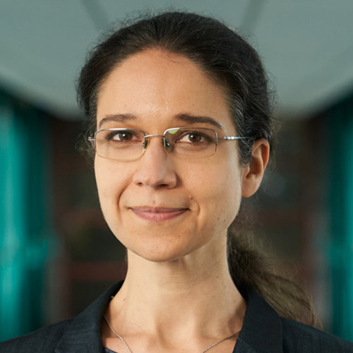
Co-senior author

Co-senior author
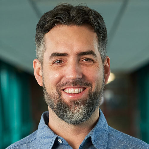
Co-senior author
Media contact
About Mass General Brigham
Mass General Brigham is an integrated academic health care system, uniting great minds to solve the hardest problems in medicine for our communities and the world. Mass General Brigham connects a full continuum of care across a system of academic medical centers, community and specialty hospitals, a health insurance plan, physician networks, community health centers, home care, and long-term care services. Mass General Brigham is a nonprofit organization committed to patient care, research, teaching, and service to the community. In addition, Mass General Brigham is one of the nation’s leading biomedical research organizations with several Harvard Medical School teaching hospitals. For more information, please visit massgeneralbrigham.org.